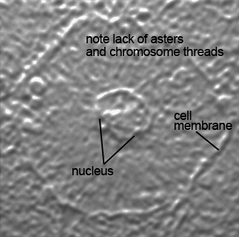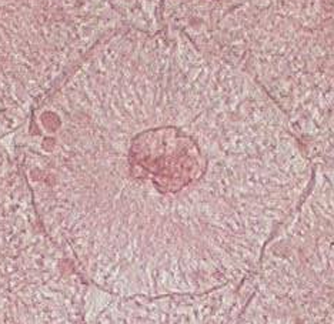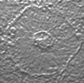
The interphase and mitotic phases listed below are shown with and without labels and are from whitefish blastula cells. The original image from which the stereolithographic (stl) file was made is also shown to give a better sense of the level of accuracy for the 3D prints which can now be use for a tactile learning experience. Digital photos of the 3D prints were made with light shining on them at an angle to show the lower regions as dark as when sunlight shines on mountains at sunset and the valleys are dark in shadow and the higher regions are still lit as this better shows the 3D nature of a print.